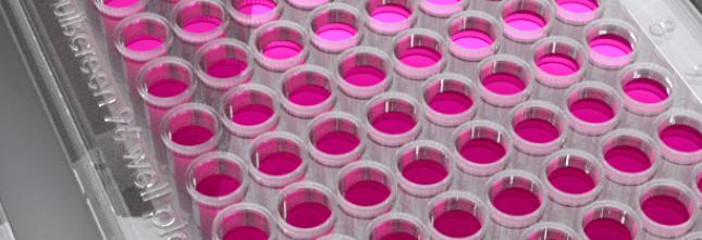
二、MICROPATTERNED MULTIWELL PLATES 96 OR 384 WELLS
For high throughput screening and high content imaging of cell-based micropatterned assays
型號:Micropatterns
聯系人:陳寶華
聯系電話:18618101725
品牌:4d cell
微圖案載片、35mm培養皿、多孔板-世聯博研quan國代理
將細胞培養在具有預定義幾何特征的微米級圖案化的基質中。當在其上進行培養時,細胞會te異地附著在圖案區域上,從而獲得例如圖案的幾何形狀。
不同的共價結合表面化學性質(圖案壽命,細胞性質):
標準抗粘聚合物
抗氧化抗粘聚合物
長期細胞培養抗粘 聚合物
不同的圖案形狀和尺寸(線,正方形,三角形,網格等)
適用于任何細胞培養底物(從培養皿到384孔板)
與高分辨率光學顯微鏡系統兼容
應用領域
細胞形狀控制和標準化,細胞定位的特征,細胞成熟和分化,遷移區域的限制等
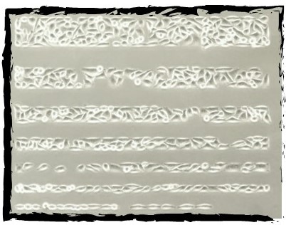
寬度為100至10 um的行中的HeLa細胞
應用示例
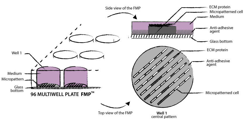
微圖案多孔板
微圖案多孔板可容納旨在組織細胞正常化的完美組織的培養底物。細胞均勻分布在膠粘劑圖案上,具有可控的幾何形狀,可實現化驗的標準化。
微圖案技術可實現2D單元幾何形狀控制。一組圖案布置在玻璃底部以容納細胞。使用抗粘劑和某些ECM蛋白(例如纖連蛋白或膠原蛋白),細胞可以粘附到微圖案所強加的形狀。單元將采用這種新環境的幾何結構。
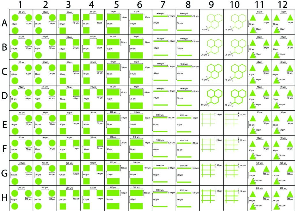
FMP micropatterning TM是一塊96孔板,其表面具有微圖案,可控制細胞的2D幾何形狀并標準化細胞測定。
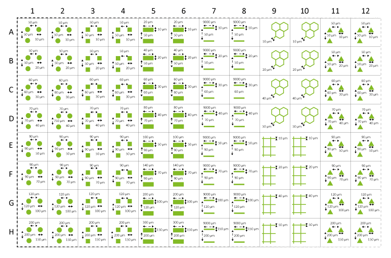
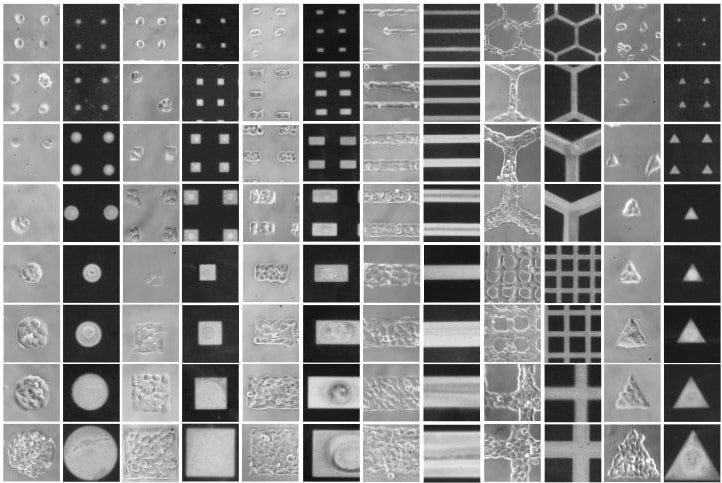
每個96孔板均具有6種標準形狀:點,正方形,矩形,三角形,直線和網格,尺寸從10到150 um。我們還提供可定制解決方案的選擇。
FMP micropatterning TM與篩選平臺的工業標準兼容。
FMP板的底部由玻璃制成,并涂有防粘聚合物以劃定圖案的邊界。該涂層具有抗氧化性,使其足以在幾天內將電池電鍍在圖案上。
標準FMP微型圖案
FMP Micropatterning TM具有廣泛的應用范圍。研究和測量:
癌癥
· 癌細胞遷移(線型)
· 像元形狀標準化
器官生理學
· 體細胞和癌細胞的遷移
· 像元形狀標準化
· 心肌搏動特性
· 神經網絡
罕見疾病
· 細胞核完整性
· 核可塑性
· 神經網絡
基礎研究
· 像元形狀標準化
· 神經元之間的標準化連接
體外生物分子模型
·
大分子的空間自組織
(微觀)
微圖案皿
微圖案化的幻燈片是印有微圖案的24毫米玻璃幻燈片。
它們已經準備好使用,您只需要將細胞鋪在玻璃基板上,并在te定的微圖案上觀察它們即可。
提供的標準形狀是圓盤,直線,正方形,矩形,三角形和網格。
您也可以要求我們為您設計的定制形狀。
二、MICROPATTERNED MULTIWELL PLATES 96 OR 384 WELLS
For high throughput screening and high content imaging of cell-based micropatterned assays
micropatterned multi-well plates hold a perfectly organized and stable culture substrate aiming for cell normalization. Evenly distributed on adhesive patterns, cells have a controlled geometry allowing standardization of the assays.
micropatterning technology enables 2D cell geometry control. A set of patterns is arranged on a glass bottom to receive cells. Using an anti-adhesive agent and some ECM proteins such as fibronectin or collagen, cells adhere and acquire the shape imposed by the micropattern.
> IMAGING OF SINGLE CELLS AND ORGANIZED CELL GROUPS
> ROBOT PIPETTE READY / SBS 96 OR 384-WELL PLATES
> HIGH-RESOLUTION IMAGING
> COMPATIBLE WITH IMAGING SOFTWARE COMMERCIALLY AVAILABLE
> MINIMAL AUTO-FLUORESCENCE
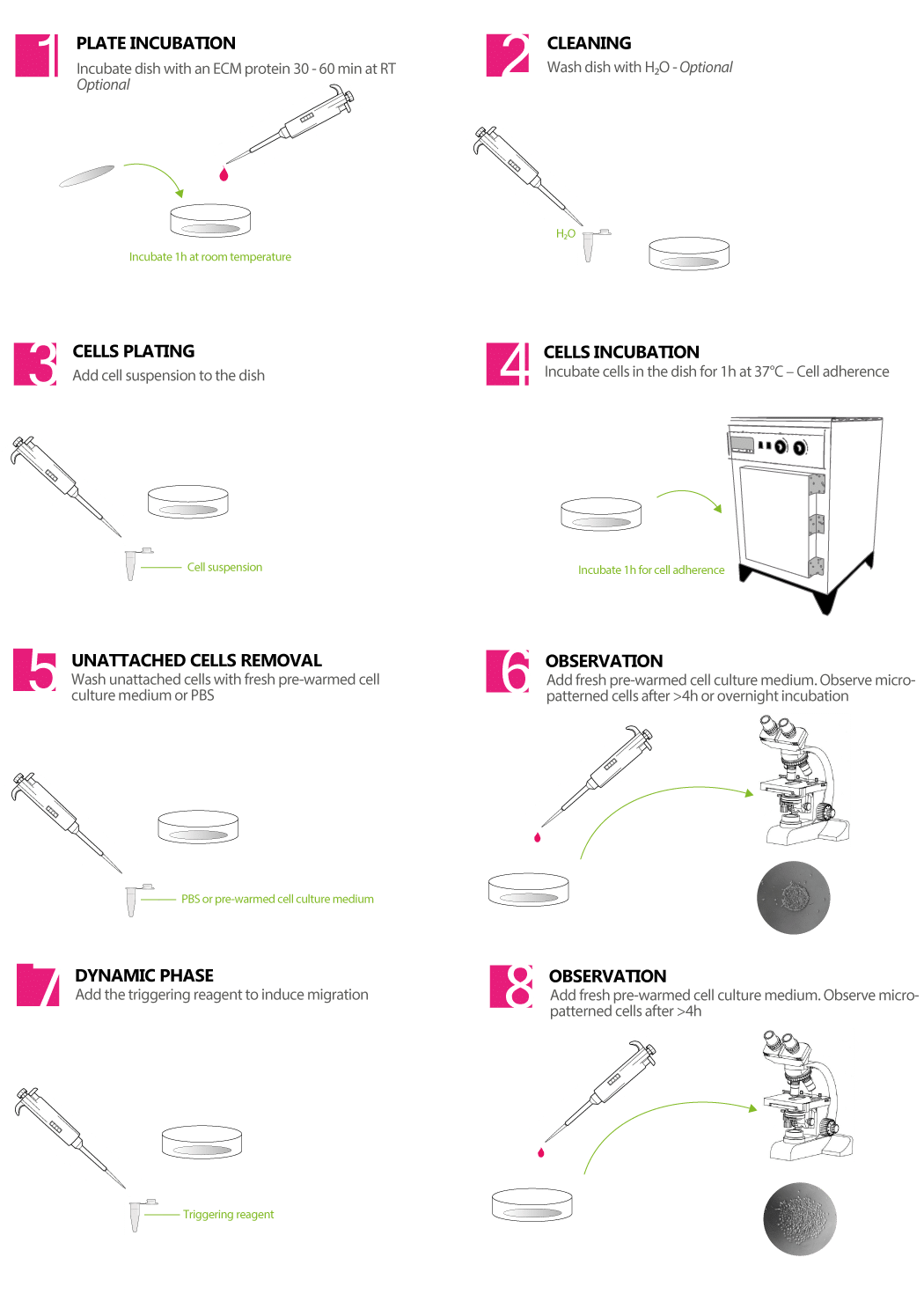
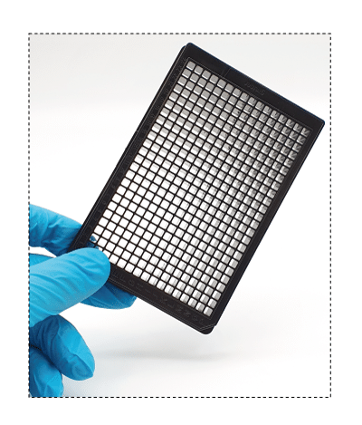
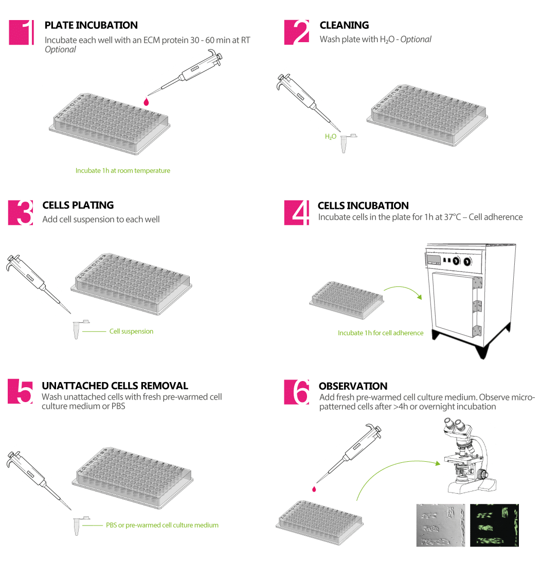
HOW TO HANDLE YOUR 96 OR 384 MICROPATTERNED WELL PLATES
FEATURES & BENEFITS
> micropatterned 96 well plates hold a patterned surface to control the 2D geometry of cells and standardize cellular assays.
> Each 96-well plate has the 6 standard shapes of : dots, squares, rectangles, triangles, lines, and grids, as well as sizes going from 10 to 150 ?m.
> We also offer the choice of a customizable solution (additional shapes and dimensions).
> The device is compatible with industrial standards for screening platforms (e.g. drug discovery).
> The bottom of the plate is made in glass and is coated with a stable anti-adhesive polymer to delimitate the borders of the patterns. This coating is resistant to oxidation, can be kept at room temperature for six months, and enables keeping the cells in patterns for several days.
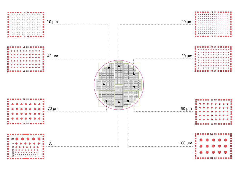
Map of the standard Micropatterned 96 well plate
DRUG SCREENING
> High throughput and high content screening
ORGAN PHYSIOLOGY & DISEASE
> Migration of somatic and cancer cells
> Cell shape standardization
> Cardiomyocyte beating properties
> Neural network and neuromuscular junction assays
> Cell nucleus integrity
> Nuclear plasticity
> Co-culture
> Wound healing
> Cellular division
> Cell-cell contact and interaction
> Cell polarization
> Frustrated phagocytosis
> Cellular membrane protrusion assays (lamellipodia and filipodia)
BIOMOLECULAR IN VITRO MODELS
> Spatial auto-organization of macromolecules and organelles (at the microscale)
三、DYNAMIC MICROPATTERNED COVERSLIPS,動態微圖案載片
Patterned slides ready to be plated with cells for cell migration assays
>即用型
HOW TO HANDLE YOUR ‘DYNAMIC’ MICROPATTERNED COVERSLIPS
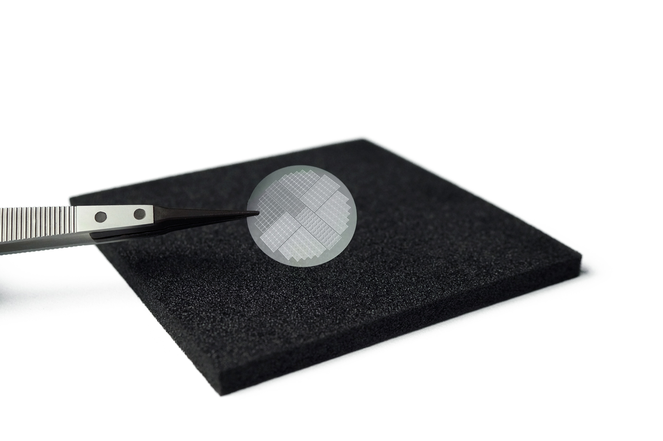
The micropatterned dishes are provided in individual sterile bags (aseptic conditions). Available in sets of 20 or 50 dishes to allow optimization oy your experimental conditions with only one kit.
Compatible with high resolution microscopy, phase contrast, immunofluorescence, etc.
The coverslips may be combined with a perfusion chamber.
MICROPATTERNS STANDARD SHAPES AVAILABLE:
As with regular micropatterned coverslips and dishes, the available shapes are:
Disks
Lines
Triangles
Squares
Rectangles
Grids
Customized shapes
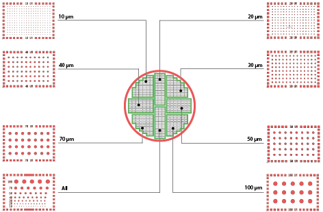
Each type of micropatterned coverslip (disks, rectangles, squares, etc.) is arranged as described in the scheme bellow (e.g.: disks).
In each standard coverslip, there are seven zones with distinct pattern sizes (ranging from 10 ?m to 100 ?m). There is an eighth zone where all the sizes are placed together.
Standard positionning of micropattern areas
A HIGHLY STABLE NON-ADHESIVE COVALENT COATING
Long term cell culture anti-adhesive polymer which is the best in terms of anti-fouling treatment and bonding to the glass slide. Observable by phase contrast microscopy. This is the best option for long term cell culture on micropatterns (more than a week of cell culture).
A HIGHLY EFFICIENT REAGENT FOR RENDERING THE COATING ADHESIVE
he reagent for switching the coating from ‘non-adhesive’ to ‘ adhesive’ and activate cell migration is shipped in aliquots of 50 ug lyophilized powder. Once dissolved in its appropriate solvent, it can be stored at -20°C.
將細胞直接鋪在帶圖案的蓋玻片上,并直接觀察
>高品質的細胞成像
通過光學透明的微圖案玻璃基板觀察細胞
>非常穩定的涂層
在防粘涂層和玻璃蓋玻片之間形成的共價鍵可在室溫下保存幾個月
>可能通過雙正交化學法制成的涂層
shou先在細胞粘附區域中將細胞圖案化,并且不粘附在這些區域的外部。 通過將一種試劑添加到基材上,可以使周圍的抗粘連區域具有粘性:這會誘導細胞遷移到新的粘連區域。